
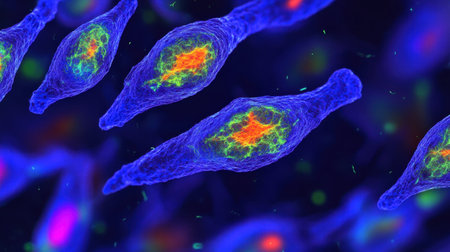
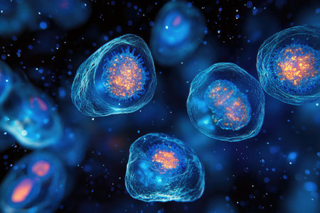
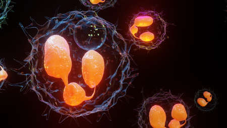
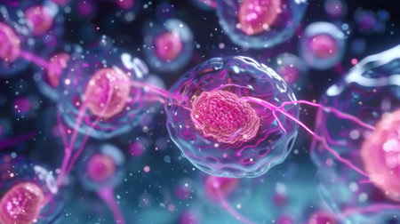
This vibrant image showcases abstract cellular structures captured through microscopy, highlighting intricate details and patterns significant in biological research and education.
Коллекция по умолчанию
Коллекция по умолчанию
Создать новую
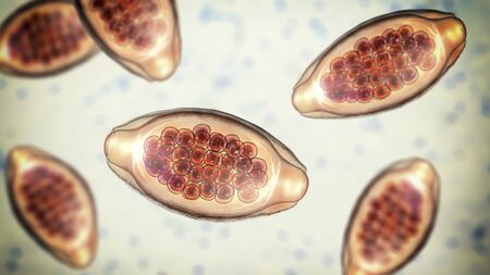
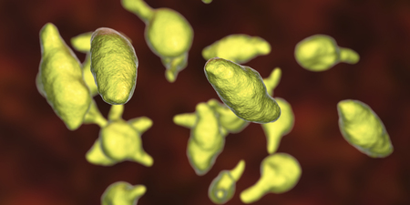
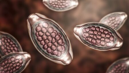
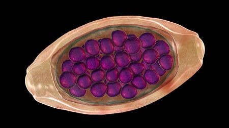
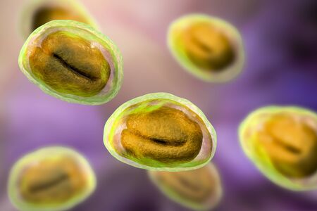
Eggs of parasitic roundworm Trichuris trichiura, or whipworm, the causative agent of trichuriasis, disease of a human large intestine, 3D illustration
Коллекция по умолчанию
Коллекция по умолчанию
Создать новую
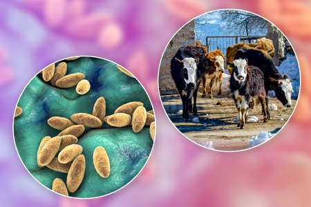
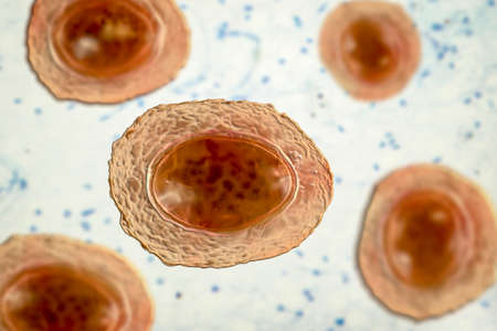
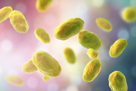
Brucellosis. 3D illustration of bacteria Brucella and photo of a cow. Brucella bacteria are transmitted to human from cattle and other animals by direct contact with ill animal or by contaminated milk
Коллекция по умолчанию
Коллекция по умолчанию
Создать новую
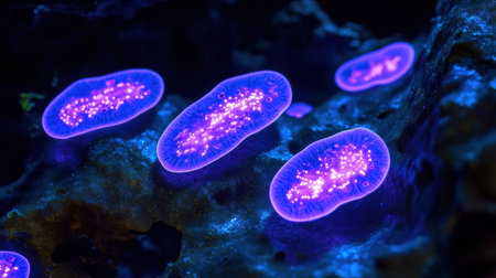
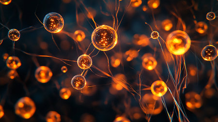
Bacteria emit a brilliant glow under UV light in a dark setting, showing vibrant colors and intricate patterns.
Коллекция по умолчанию
Коллекция по умолчанию
Создать новую
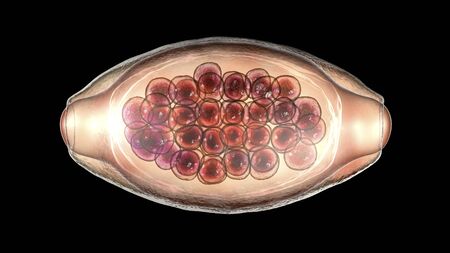
Egg of parasitic roundworm Trichuris trichiura, or whipworm, the causative agent of trichuriasis, disease of a human large intestine, 3D illustration
Коллекция по умолчанию
Коллекция по умолчанию
Создать новую
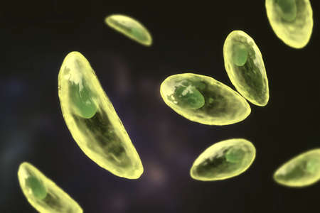
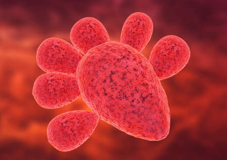
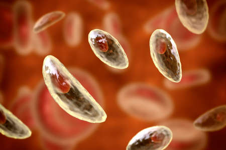
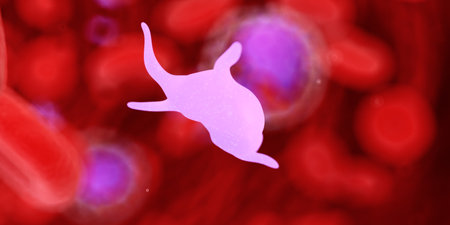
Parasitic protozoans Toxoplasma gondii, the causative agent of toxoplasmosis in tachyzoite stage, 3D illustration
Коллекция по умолчанию
Коллекция по умолчанию
Создать новую
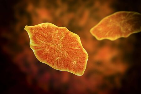
Fasciola hepatica, or liver fluke, 3D illustration. A parasitic trematode worm that causes fasciolosis, an infection of liver
Коллекция по умолчанию
Коллекция по умолчанию
Создать новую
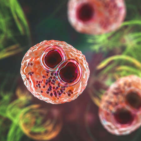
Cytomegalovirus CMV in a human cell, owl's eye inclusion in nucleus, multinucleated cell, 3D illustration. It is herpes virus, causes diseases in fetus, organ transplant patients, HIV infected people
Коллекция по умолчанию
Коллекция по умолчанию
Создать новую
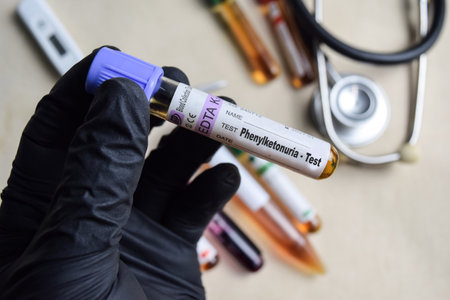
Phenylketonuria - Test with blood sample in black gloved hands. healthcare or Medical concept
Коллекция по умолчанию
Коллекция по умолчанию
Создать новую
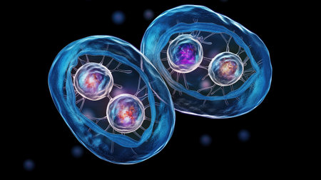
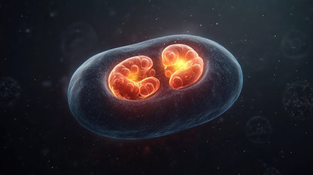
Cell division process, micro
Коллекция по умолчанию
Коллекция по умолчанию
Создать новую
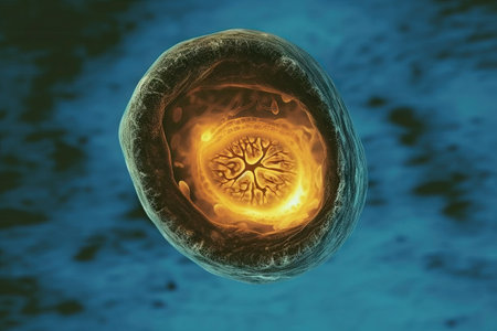
Human egg cell, 3D illustration closeup
Коллекция по умолчанию
Коллекция по умолчанию
Создать новую

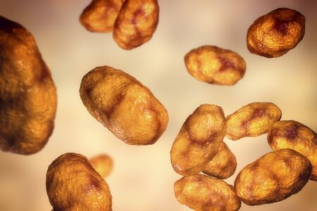
Cancer Cell in human showing abnormal cells.Medical science background concept.
Коллекция по умолчанию
Коллекция по умолчанию
Создать новую
Haemophilus influenzae bacteria, 3D illustration. Gram-negative coccobacilli which cause infections mainly in children, pneumonia, otitis, meninitis and other
Коллекция по умолчанию
Коллекция по умолчанию
Создать новую

Realistic molecule bacterium illustration graphics 3d microbe
Коллекция по умолчанию
Коллекция по умолчанию
Создать новую
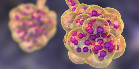
Coronavirus pneumonia, alveoli filled with Covid-19
Коллекция по умолчанию
Коллекция по умолчанию
Создать новую

Slide sputum AFB.
Коллекция по умолчанию
Коллекция по умолчанию
Создать новую
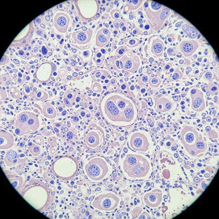
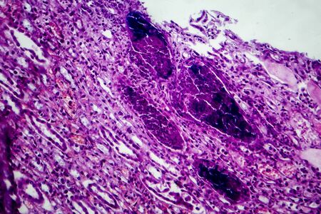
Thyroid follicular carcinoma, light micrograph, photo under microscope
Коллекция по умолчанию
Коллекция по умолчанию
Создать новую
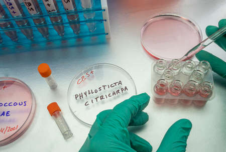
Samples of a tiny fungus in oranges called Phyllosticta citricarpa or black spot, it is a pest especially present in South Africa, petri dish contains residues, conceptual image
Коллекция по умолчанию
Коллекция по умолчанию
Создать новую
Close-up of a single biological cell with glowing edges and complex internal structures on a dark background. Copy space.
Коллекция по умолчанию
Коллекция по умолчанию
Создать новую

Blue spots from the dye in the white tub dissolves in water
Коллекция по умолчанию
Коллекция по умолчанию
Создать новую
3d illustration of transparent glowing human cells with visible organelles, nucleus and DNA
Коллекция по умолчанию
Коллекция по умолчанию
Создать новую
Bacteria Mycoplasma genitalium, 3D illustration. The causative agent of sexually transmitted infections and infertility
Коллекция по умолчанию
Коллекция по умолчанию
Создать новую

Coronavirus pneumonia, alveoli filled with Covid-19
Коллекция по умолчанию
Коллекция по умолчанию
Создать новую
Ascaris lumbricoides, a large roundworm, unfertilized egg, 3D illustration
Коллекция по умолчанию
Коллекция по умолчанию
Создать новую
Microscopic view of human neurons. Neuron cells. 3D rendering
Коллекция по умолчанию
Коллекция по умолчанию
Создать новую
3D illustration of Phagocytosis. Neutrophe that uses its plasma membrane to engulf bacteria. From endocytosis to exocytosis. Digestion process in phagocytes. immune system, 3d render
Коллекция по умолчанию
Коллекция по умолчанию
Создать новую

Medical research sample in laboratory
Коллекция по умолчанию
Коллекция по умолчанию
Создать новую
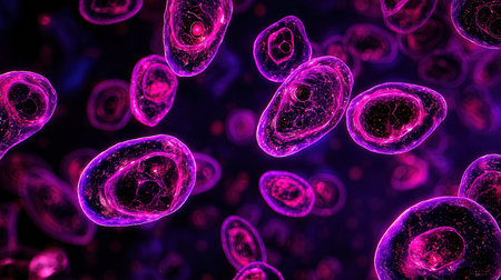
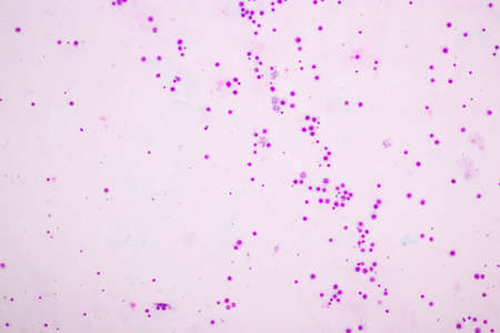
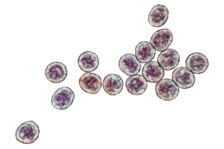
Microscopic purple cell cluster pattern abstract
Коллекция по умолчанию
Коллекция по умолчанию
Создать новую

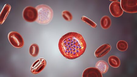
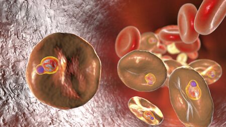
The malaria-infected red blood cells. 3D illustration showing malaria parasite Plasmodium falciparum in schizont stage inside red blood cells, the causative agent of tropical malaria
Коллекция по умолчанию
Коллекция по умолчанию
Создать новую

Eggs of parasitic roundworm Trichuris trichiura, or whipworm, the causative agent of trichuriasis, disease of a human large intestine, 3D illustration
Коллекция по умолчанию
Коллекция по умолчанию
Создать новую

Abstract macro image of particles looking like bacteria, macro shot, microbiology theme
Коллекция по умолчанию
Коллекция по умолчанию
Создать новую
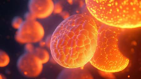
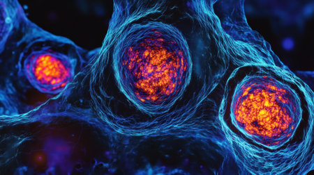
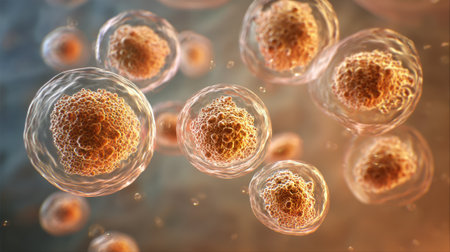
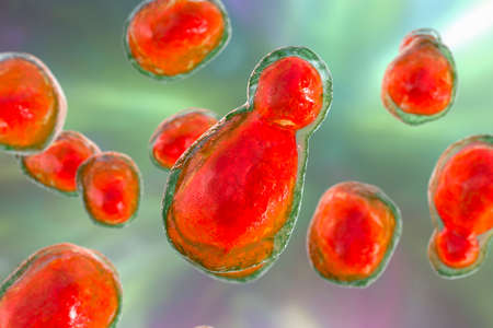
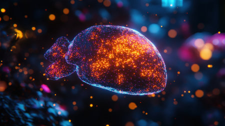
This vibrant abstract image showcases glowing microbial cells in shades of orange and yellow, highlighting their unique textures and forms, perfect for scientific themes.
Коллекция по умолчанию
Коллекция по умолчанию
Создать новую

Claustrophobia. The text inscription on the plate of the banner. This is the fear of a closed space, caused by situations or stimuli.
Коллекция по умолчанию
Коллекция по умолчанию
Создать новую
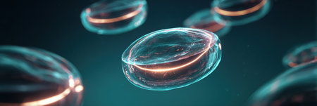
Translucent spheres hover in a dark space, exhibiting glowing edges and smooth curves, creating a captivating and futuristic ambiance.
Коллекция по умолчанию
Коллекция по умолчанию
Создать новую
Under ultraviolet light, bacteria emit a vibrant glow, revealing their bioluminescent properties in a dark, captivating setting.
Коллекция по умолчанию
Коллекция по умолчанию
Создать новую

Parasitic protozoans Toxoplasma gondii, the causative agent of toxoplasmosis in tachyzoite stage, 3D illustration
Коллекция по умолчанию
Коллекция по умолчанию
Создать новую
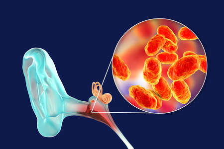
Otitis media, inflammatory disease of the middle ear
Коллекция по умолчанию
Коллекция по умолчанию
Создать новую
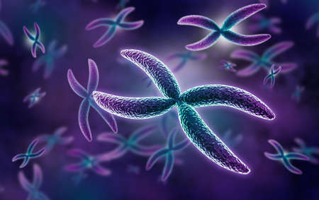
Chromosomes Human under the microscope for education.
Коллекция по умолчанию
Коллекция по умолчанию
Создать новую

This vibrant microscopic image showcases colorful cells illuminated by fluorescent light. Ideal for scientific research, biology studies, or educational purposes, this image captures the beauty of cellular structures.
Коллекция по умолчанию
Коллекция по умолчанию
Создать новую
Chromosomes Human under the microscope for education.
Коллекция по умолчанию
Коллекция по умолчанию
Создать новую
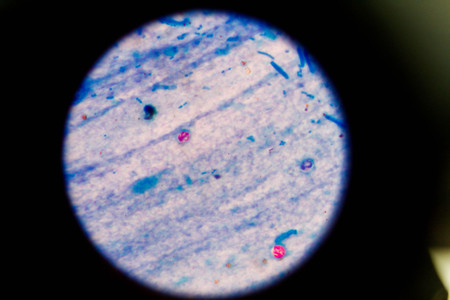
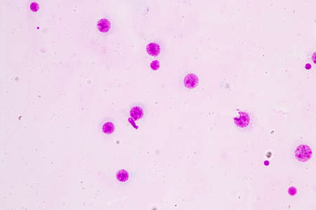
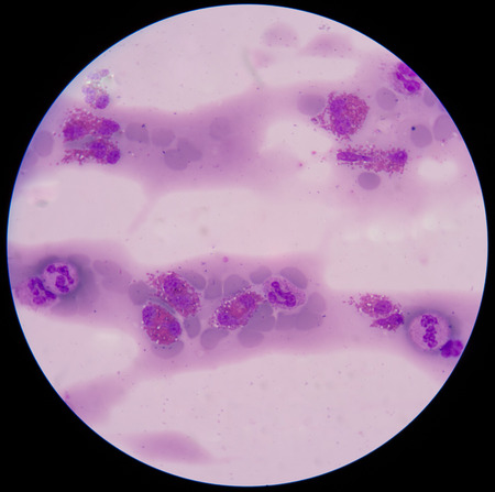
stool specimen shows Cryptosporidium eggs in red. They can cause diarrhea and more serious problems in children and adults who immune systems are suppressed.
Коллекция по умолчанию
Коллекция по умолчанию
Создать новую

Abnormal cells pink cells with microscope.Medical science background concept.
Коллекция по умолчанию
Коллекция по умолчанию
Создать новую
Histopathology of human liver under microscope view for medical education.
Коллекция по умолчанию
Коллекция по умолчанию
Создать новую
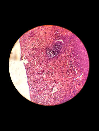
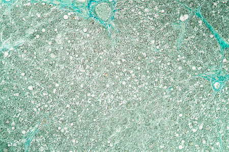
Biological histological fixed colored preparation of the spleen - a secondary organ of the immune system
Коллекция по умолчанию
Коллекция по умолчанию
Создать новую

Cytomegalovirus CMV in a human cell, owl's eye inclusion in nucleus, multinucleated cell, 3D illustration. It is herpes virus, causes diseases in fetus, organ transplant patients, HIV infected people
Коллекция по умолчанию
Коллекция по умолчанию
Создать новую
A closeup examination of a cultured cell on a nanoengineered substrate with bright staining revealing cellular morphology and interaction with the surface at the nanoscale
Коллекция по умолчанию
Коллекция по умолчанию
Создать новую
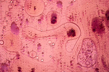
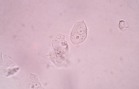
Soothing Pink Abstract background with little impurities
Коллекция по умолчанию
Коллекция по умолчанию
Создать новую
Paracoccidioides fungus is the cause of paracoccidioidomycosis is a systemic mycosis that occurs in humans. 3D rendering
Коллекция по умолчанию
Коллекция по умолчанию
Создать новую
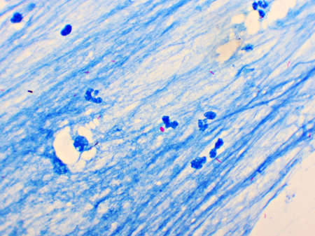
Mycobacterium tuberculosis positive (small red rod) in sputum smear, acid-fast stain, analyze by microscope 1000x
Коллекция по умолчанию
Коллекция по умолчанию
Создать новую
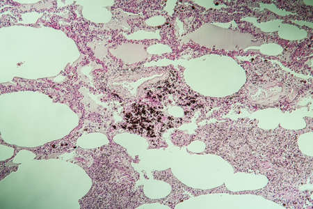
Lung tissue as dust lung under the microscope 100x
Коллекция по умолчанию
Коллекция по умолчанию
Создать новую
cell find with microscope.
Коллекция по умолчанию
Коллекция по умолчанию
Создать новую
bacteria under the microscope background, background bacteria
Коллекция по умолчанию
Коллекция по умолчанию
Создать новую

brucella bacteria is a pathogen that causes brucellosis zoonosis. 3D rendering
Коллекция по умолчанию
Коллекция по умолчанию
Создать новую
Pancreas cancer cells, light micrograph for medical education.
Коллекция по умолчанию
Коллекция по умолчанию
Создать новую
Parasitic protozoans Toxoplasma gondii, the causative agent of toxoplasmosis in tachyzoite stage, 3D illustration
Коллекция по умолчанию
Коллекция по умолчанию
Создать новую
Eggs of parasitic roundworm Trichuris trichiura, or whipworm, the causative agent of trichuriasis, disease of a human large intestine, 3D illustration
Коллекция по умолчанию
Коллекция по умолчанию
Создать новую
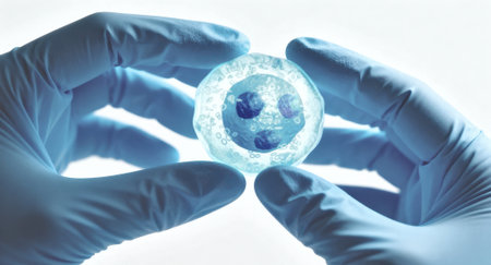
Gloved hands hold a petri dish containing a cultured cell cluster under cool blue backlighting, suggesting laboratory research and biomedical analysis with sterile technique and visible liquid medium ready for examination
Коллекция по умолчанию
Коллекция по умолчанию
Создать новую
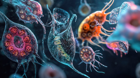
Dive into a stunning microscopic view showcasing vibrant microorganisms in an aquatic setting. This image reveals the intricacies of diverse life forms through scientific observation.
Коллекция по умолчанию
Коллекция по умолчанию
Создать новую
blood films for Malaria parasite.
Коллекция по умолчанию
Коллекция по умолчанию
Создать новую
Yersinia enterocolitica bacteria, 3D illustration. Gram-negative bacteria of Enterobacteriaceae family, the causative agent of Yersiniosis
Коллекция по умолчанию
Коллекция по умолчанию
Создать новую

Hand of a doctor with blue glove on it holding a bottle for samples.
Коллекция по умолчанию
Коллекция по умолчанию
Создать новую

medical bacteria illustration of the clostridium
Коллекция по умолчанию
Коллекция по умолчанию
Создать новую
Egg of parasitic roundworm Trichuris trichiura, or whipworm, the causative agent of trichuriasis, disease of a human large intestine, 3D illustration
Коллекция по умолчанию
Коллекция по умолчанию
Создать новую
A close-up of healthy cells magnified under a light microscope. The cells exhibit detailed structures and activities, showing the complexity of life at a microscopic level.
Коллекция по умолчанию
Коллекция по умолчанию
Создать новую
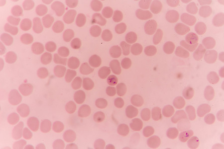
plasmodium falciparum ring form state , multiple in fection of red blood cells
Коллекция по умолчанию
Коллекция по умолчанию
Создать новую
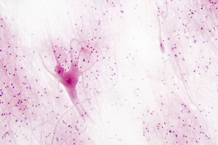
Motor nerves in spinal tissue.
Коллекция по умолчанию
Коллекция по умолчанию
Создать новую
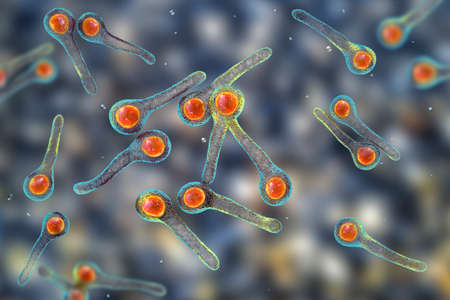
Clostridium tetani bacteria, the causative agent of tetanus, 3D illustration
Коллекция по умолчанию
Коллекция по умолчанию
Создать новую

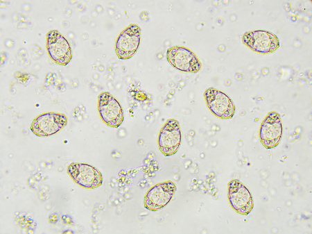
The human whipworm (Trichuris trichiura or Trichocephalus trichiuris) is a round worm (a type of helminth) that causes trichuriasis (a type of helminthiasis which is one of the neglected tropical diseases) when it infects a human large intestine.
Коллекция по умолчанию
Коллекция по умолчанию
Создать новую
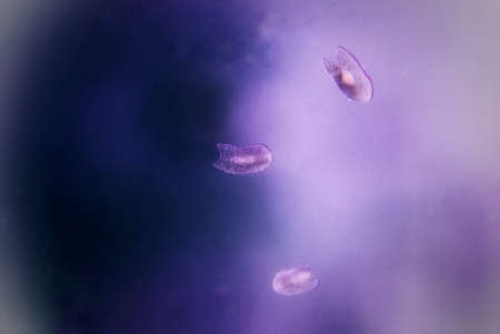
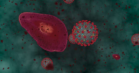
Rare image of Ghost flatworm - Maricola (Planarian) triclad flatworms in reef aquarium glass
Коллекция по умолчанию
Коллекция по умолчанию
Создать новую
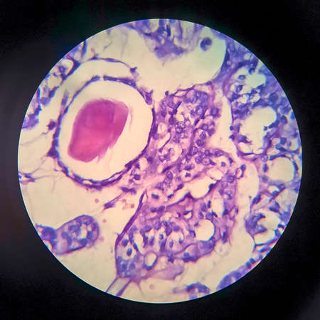
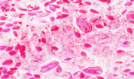
Eggs of Opisthorchis viverrini in stool
Коллекция по умолчанию
Коллекция по умолчанию
Создать новую
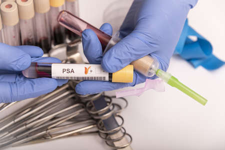
Blood sample with abnormal high PSA test result.
Коллекция по умолчанию
Коллекция по умолчанию
Создать новую
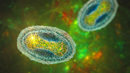
Molluscum contagiosum virus, 3D illustration. A virus from Poxvirus family, causes skin infection with numerous small raised lesions
Коллекция по умолчанию
Коллекция по умолчанию
Создать новую
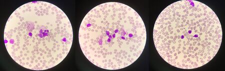
Immature white blood cells in leukemia.Science concept.
Коллекция по умолчанию
Коллекция по умолчанию
Создать новую
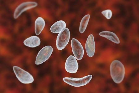
Parasitic protozoans Toxoplasma gondii which cause toxoplasmosis in tachyzoite stage, 3D illustration
Коллекция по умолчанию
Коллекция по умолчанию
Создать новую
The malaria-infected red blood cells. 3D illustration showing malaria parasite Plasmodium falciparum in schizont stage inside red blood cells, the causative agent of tropical malaria
Коллекция по умолчанию
Коллекция по умолчанию
Создать новую
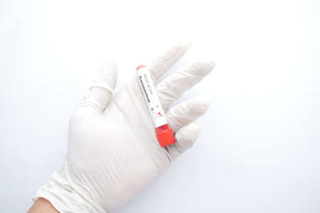
hand in medical gloves holding blood test tube on white background
Коллекция по умолчанию
Коллекция по умолчанию
Создать новую

Neurons in the brain from microscope.(Soft focus)
Коллекция по умолчанию
Коллекция по умолчанию
Создать новую

Mycobacterium tuberculosis undermicroscope 100x /AFB POSITIVE 3+
Коллекция по умолчанию
Коллекция по умолчанию
Создать новую
Pathogenic yeast fungus Cryptococcus neoformans that causes cryptococcal meningoencephalitis and lung disease in immunocompromised patients with AIDS
Коллекция по умолчанию
Коллекция по умолчанию
Создать новую
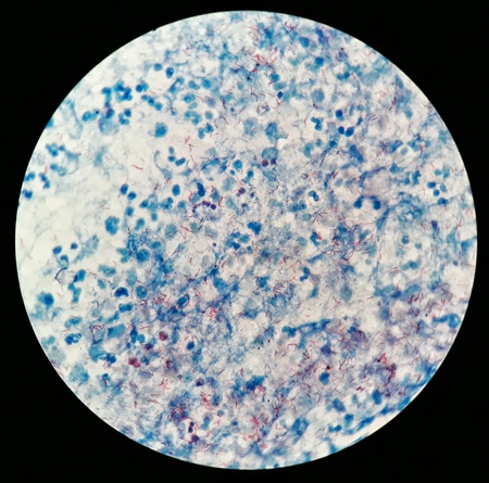
Smear of Positive Acid Fast Bacilli AFB stained for MTB, under 100X light microscope.
Коллекция по умолчанию
Коллекция по умолчанию
Создать новую

Smear of Acid-Fast bacilli AFB stained from sputum specimen, under 100X light microscope.
Коллекция по умолчанию
Коллекция по умолчанию
Создать новую
3d rendered medically accurate illustration of cells
Коллекция по умолчанию
Коллекция по умолчанию
Создать новую
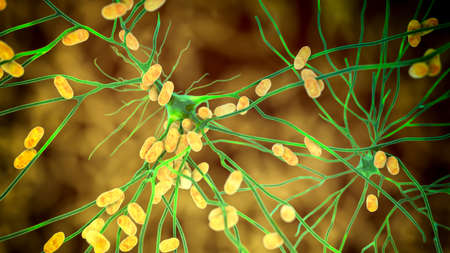
Bacteria infecting neuron, brain cell, 3D illustration. Conceptual illustration of bacterial encephalitis, meningitis, bacterial infection of brain tissue
Коллекция по умолчанию
Коллекция по умолчанию
Создать новую
chromosome in digital background
Коллекция по умолчанию
Коллекция по умолчанию
Создать новую

Euglena is a genus of single-celled flagellate Eukaryotes under microscopic view for education.
Коллекция по умолчанию
Коллекция по умолчанию
Создать новую
Rat liver 100x tissue section
Коллекция по умолчанию
Коллекция по умолчанию
Создать новую
Human cell under microscope.
Коллекция по умолчанию
Коллекция по умолчанию
Создать новую
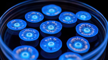
This captivating image showcases vibrant blue jellyfish in a petri dish against a deep black background, illustrating the beauty of marine life. Perfect for scientific and educational use.
Коллекция по умолчанию
Коллекция по умолчанию
Создать новую
Eggs of helminths Enterobius vermicularis containing worm larva. Threadworm which cause enterobiasis, 3D illustration
Коллекция по умолчанию
Коллекция по умолчанию
Создать новую
blood smear is often used as a follow-up test to abnormal results on a complete blood count (CBC) to evaluate the different types of blood cells.
Коллекция по умолчанию
Коллекция по умолчанию
Создать новую
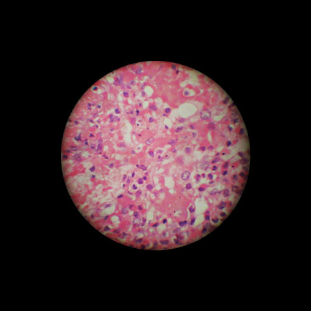
image through the microscope. zoom x 40
Коллекция по умолчанию
Коллекция по умолчанию
Создать новую
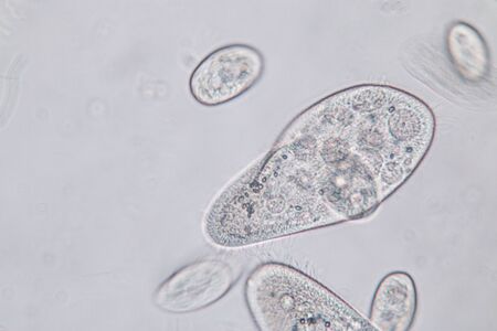
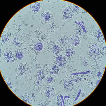
Paramecium caudatum is a genus of unicellular ciliated protozoan and Bacterium under the microscope.
Коллекция по умолчанию
Коллекция по умолчанию
Создать новую
Hight concentration Coronavirus disease Covid-19. Animation group of viruses and Red blood cells close up under the microscope. 3D rendering 3D illustration
Коллекция по умолчанию
Коллекция по умолчанию
Создать новую
crystal cell
Коллекция по умолчанию
Коллекция по умолчанию
Создать новую
Bacteria methicillin-resistant Staphylococcus aureus MRSA, multidrug resistant bacteria, 3D illustration
Коллекция по умолчанию
Коллекция по умолчанию
Создать новую

3D illustration of abstract light background with bokeh defocused lights
Коллекция по умолчанию
Коллекция по умолчанию
Создать новую
Abstract heart, glowing particles, dark background, medical research
Коллекция по умолчанию
Коллекция по умолчанию
Создать новую
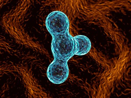
Babesia parasites inside red blood cell, the causative agent of babesiosis. 3D illustration showing classic tetrad-forms of Babesia merozoites so-called Maltese cross formation
Коллекция по умолчанию
Коллекция по умолчанию
Создать новую

Blastomyces dermatitidis fungi, the causative agent of the disease blastomycosis affecting lungs, more rarely skin, bones, other organs, 3D illustration. Yeast form
Коллекция по умолчанию
Коллекция по умолчанию
Создать новую
Acute pyelonephritis, light micrograph, photo under microscope
Коллекция по умолчанию
Коллекция по умолчанию
Создать новую
Legion-Media
Создайте свои проекты на основе качественных стоковых фотографий и видео.


Copyright © Legion-Media.